Example of Substances That Use Facilitated Diffusion in Cells
Facilitated transport
Facilitated diffusion is a process by which molecules are transported across the plasma membrane with the help of membrane proteins.
Learning Objectives
Explain why and how passive transport occurs
Key Takeaways
Key Points
- A concentration gradient exists that would allow ions and polar molecules to diffuse into the cell, but these materials are repelled by the hydrophobic parts of the cell membrane.
- Facilitated diffusion uses integral membrane proteins to move polar or charged substances across the hydrophobic regions of the membrane.
- Channel proteins can aid in the facilitated diffusion of substances by forming a hydrophilic passage through the plasma membrane through which polar and charged substances can pass.
- Channel proteins can be open at all times, constantly allowing a particular substance into or out of the cell, depending on the concentration gradient; or they can be gated and can only be opened by a particular biological signal.
- Carrier proteins aid in facilitated diffusion by binding a particular substance, then altering their shape to bring that substance into or out of the cell.
Key Terms
- facilitated diffusion: The spontaneous passage of molecules or ions across a biological membrane passing through specific transmembrane integral proteins.
- membrane protein: Proteins that are attached to, or associated with the membrane of a cell or an organelle.
Facilitated transport
Facilitated transport is a type of passive transport. Unlike simple diffusion where materials pass through a membrane without the help of proteins, in facilitated transport, also called facilitated diffusion, materials diffuse across the plasma membrane with the help of membrane proteins. A concentration gradient exists that would allow these materials to diffuse into the cell without expending cellular energy. However, these materials are ions or polar molecules that are repelled by the hydrophobic parts of the cell membrane. Facilitated transport proteins shield these materials from the repulsive force of the membrane, allowing them to diffuse into the cell.
The material being transported is first attached to protein or glycoprotein receptors on the exterior surface of the plasma membrane. This allows the material that is needed by the cell to be removed from the extracellular fluid. The substances are then passed to specific integral proteins that facilitate their passage. Some of these integral proteins are collections of beta-pleated sheets that form a channel through the phospholipid bilayer. Others are carrier proteins which bind with the substance and aid its diffusion through the membrane.
Channels
The integral proteins involved in facilitated transport are collectively referred to as transport proteins; they function as either channels for the material or carriers. In both cases, they are transmembrane proteins. Channels are specific for the substance that is being transported. Channel proteins have hydrophilic domains exposed to the intracellular and extracellular fluids; they additionally have a hydrophilic channel through their core that provides a hydrated opening through the membrane layers. Passage through the channel allows polar compounds to avoid the nonpolar central layer of the plasma membrane that would otherwise slow or prevent their entry into the cell. Aquaporins are channel proteins that allow water to pass through the membrane at a very high rate.

Channel Proteins in Facilitated Transport: Facilitated transport moves substances down their concentration gradients. They may cross the plasma membrane with the aid of channel proteins.
Channel proteins are either open at all times or they are "gated," which controls the opening of the channel. The attachment of a particular ion to the channel protein may control the opening or other mechanisms or substances may be involved. In some tissues, sodium and chloride ions pass freely through open channels, whereas in other tissues, a gate must be opened to allow passage. An example of this occurs in the kidney, where both forms of channels are found in different parts of the renal tubules. Cells involved in the transmission of electrical impulses, such as nerve and muscle cells, have gated channels for sodium, potassium, and calcium in their membranes. Opening and closing of these channels changes the relative concentrations on opposing sides of the membrane of these ions, resulting in the facilitation of electrical transmission along membranes (in the case of nerve cells) or in muscle contraction (in the case of muscle cells).
Carrier Proteins
Another type of protein embedded in the plasma membrane is a carrier protein. This protein binds a substance and, in doing so, triggers a change of its own shape, moving the bound molecule from the outside of the cell to its interior; depending on the gradient, the material may move in the opposite direction. Carrier proteins are typically specific for a single substance. This adds to the overall selectivity of the plasma membrane. The exact mechanism for the change of shape is poorly understood. Proteins can change shape when their hydrogen bonds are affected, but this may not fully explain this mechanism. Each carrier protein is specific to one substance, and there are a finite number of these proteins in any membrane. This can cause problems in transporting enough of the material for the cell to function properly.
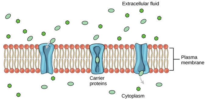
Carrier Proteins: Some substances are able to move down their concentration gradient across the plasma membrane with the aid of carrier proteins. Carrier proteins change shape as they move molecules across the membrane.
An example of this process occurs in the kidney. Glucose, water, salts, ions, and amino acids needed by the body are filtered in one part of the kidney. This filtrate, which includes glucose, is then reabsorbed in another part of the kidney. Because there are only a finite number of carrier proteins for glucose, if more glucose is present than the proteins can handle, the excess is not transported; it is excreted from the body in the urine. In a diabetic individual, this is described as "spilling glucose into the urine." A different group of carrier proteins called glucose transport proteins, or GLUTs, are involved in transporting glucose and other hexose sugars through plasma membranes within the body.
Channel and carrier proteins transport material at different rates. Channel proteins transport much more quickly than do carrier proteins. Channel proteins facilitate diffusion at a rate of tens of millions of molecules per second, whereas carrier proteins work at a rate of a thousand to a million molecules per second.
Primary Active Transport
The sodium-potassium pump maintains the electrochemical gradient of living cells by moving sodium in and potassium out of the cell.
Learning Objectives
Describe how a cell moves sodium and potassium out of and into the cell against its electrochemical gradient
Key Takeaways
Key Points
- The sodium-potassium pump moves K+ into the cell while moving Na+ at a ratio of three Na+ for every two K+ ions.
- When the sodium-potassium- ATPase enzyme points into the cell, it has a high affinity for sodium ions and binds three of them, hydrolyzing ATP and changing shape.
- As the enzyme changes shape, it reorients itself towards the outside of the cell, and the three sodium ions are released.
- The enzyme's new shape allows two potassium to bind and the phosphate group to detach, and the carrier protein repositions itself towards the interior of the cell.
- The enzyme changes shape again, releasing the potassium ions into the cell.
- After potassium is released into the cell, the enzyme binds three sodium ions, which starts the process over again.
Key Terms
- electrogenic pump: An ion pump that generates a net charge flow as a result of its activity.
- Na+-K+ ATPase: An enzyme located in the plasma membrane of all animal cells that pumps sodium out of cells while pumping potassium into cells.
Primary Active Transport
The primary active transport that functions with the active transport of sodium and potassium allows secondary active transport to occur. The secondary transport method is still considered active because it depends on the use of energy as does primary transport.

Active Transport of Sodium and Potassium: Primary active transport moves ions across a membrane, creating an electrochemical gradient (electrogenic transport).
One of the most important pumps in animals cells is the sodium-potassium pump ( Na+-K+ ATPase ), which maintains the electrochemical gradient (and the correct concentrations of Na+ and K+) in living cells. The sodium-potassium pump moves two K+ into the cell while moving three Na+ out of the cell. The Na+-K+ ATPase exists in two forms, depending on its orientation to the interior or exterior of the cell and its affinity for either sodium or potassium ions. The process consists of the following six steps:
- With the enzyme oriented towards the interior of the cell, the carrier has a high affinity for sodium ions. Three sodium ions bind to the protein.
- ATP is hydrolyzed by the protein carrier, and a low-energy phosphate group attaches to it.
- As a result, the carrier changes shape and re-orients itself towards the exterior of the membrane. The protein's affinity for sodium decreases, and the three sodium ions leave the carrier.
- The shape change increases the carrier's affinity for potassium ions, and two such ions attach to the protein. Subsequently, the low-energy phosphate group detaches from the carrier.
- With the phosphate group removed and potassium ions attached, the carrier protein repositions itself towards the interior of the cell.
- The carrier protein, in its new configuration, has a decreased affinity for potassium, and the two ions are released into the cytoplasm. The protein now has a higher affinity for sodium ions, and the process starts again.
Several things have happened as a result of this process. At this point, there are more sodium ions outside of the cell than inside and more potassium ions inside than out. For every three ions of sodium that move out, two ions of potassium move in. This results in the interior being slightly more negative relative to the exterior. This difference in charge is important in creating the conditions necessary for the secondary process. The sodium-potassium pump is, therefore, an electrogenic pump (a pump that creates a charge imbalance), creating an electrical imbalance across the membrane and contributing to the membrane potential.
ABC Transporters
ABC transporters are a protein superfamily that all have an ATP binding cassette and transport substances across membranes.
Learning Objectives
Summarize the function of the three major ABC transporter categories: in prokaryotes, in gram-negative bacteria and the subgroup of ABC proteins
Key Takeaways
Key Points
- ABC transporters use the energy of ATP hydrolysis to transport substrates across cell membranes.
- Bacterial ABC transporters are essential in cell viability, virulence, and pathogenicity.
- The substrates that can be transported include ions, amino acids, peptides, sugars, and other molecules that are mostly hydrophilic.
Key Terms
- membrane: A flexible enclosing or separating tissue forming a plane or film and separating two environments (usually in a plant or animal).
- hydrolysis: A chemical process of decomposition involving the splitting of a bond and the addition of the hydrogen cation and the hydroxide anion of water.
- ATP-binding cassette (ABC) domain: The ATP-binding cassette (ABC) family is a group of proteins which bind and hydrolyse ATP in order to transport substances across cellular membranes.
ATP-binding cassette transporters (ABC-transporters) are members of a protein superfamily that is one of the largest and most ancient families with representatives in all extant phyla from prokaryotes to humans.
ABC transporters are transmembrane proteins that utilize the energy of adenosine triphosphate (ATP) hydrolysis to carry out certain biological processes including translocation of various substrates across membranes and non-transport-related processes such as translation of RNA and DNA repair. They transport a wide variety of substrates across extra- and intracellular membranes, including metabolic products, lipids and sterols, and drugs. Proteins are classified as ABC transporters based on the sequence and organization of their ATP-binding cassette (ABC) domain(s).
ABC transporters are involved in tumor resistance, cystic fibrosis and a range of other inherited human diseases along with both bacterial (prokaryotic) and eukaryotic (including human) development of resistance to multiple drugs. Bacterial ABC transporters are essential in cell viability, virulence, and pathogenicity.
ABC transporters are divided into three main functional categories. In prokaryotes, importers mediate the uptake of nutrients into the cell. The substrates that can be transported include ions, amino acids, peptides, sugars, and other molecules that are mostly hydrophilic. The membrane-spanning region of the ABC transporter protects hydrophilic substrates from the lipids of the membrane bilayer thus providing a pathway across the cell membrane. In gram-negative bacteria, exporters transport lipids and some polysaccharides from the cytoplasm to the periplasm. Eukaryotes do not possess any importers. Exporters or effluxers, which are both present in prokaryotes and eukaryotes, function as pumps that extrude toxins and drugs out of the cell. The third subgroup of ABC proteins do not function as transporters, but rather are involved in translation and DNA repair processes.

Mechanism of ABC transport: Proposed mechanism of transport for ABC importers. This alternating-access model was based on the crystal structures of ModBC-A
In bacterial efflux systems, certain substances that need to be extruded from the cell include surface components of the bacterial cell (e.g. capsular polysaccharides, lipopolysaccharides, and teichoic acid), proteins involved in bacterial pathogenesis (e.g. hemolysis, heme-binding protein, and alkaline protease), heme, hydrolytic enzymes, S-layer proteins, competence factors, toxins, antibiotics, bacteriocins, peptide antibiotics, drugs and siderophores. They also play important roles in biosynthetic pathways, including extracellular polysaccharide biosynthesis and cytochrome biogenesis.
Siderophores
Siderophores are classified by which ligands they use to chelate the ferric iron, including the catecholates, hydroxamates, and carboxylates.
Learning Objectives
Describe the function and variety of siderophores
Key Takeaways
Key Points
- Siderophores are important for some pathogenic bacteria for their acquisition of iron. Many siderophores are nonribosomal peptides, although several are biosynthesised independently.
- The wide variety of siderophores may be due to evolutionary pressures placed on microbes to produce structurally different siderophores which cannot be transported by other microbes' specific active transport systems, or in the case of pathogens deactivated by the host organism.
- Microbes release siderophores to scavenge iron from these mineral phases by formation of soluble Fe3+ complexes that can be taken up by active transport mechanisms.
Key Terms
- siderophores: Sidereophores are small, high-affinity iron chelating compounds secreted by microorganisms such as bacteria and fungi, and also grasses. Siderophores are amongst the strongest soluble Fe3+ binding agents known.
Iron is essential for almost all living organisms as it is involved in a wide variety of important metabolic processes. However, iron is not always readily available; therefore, microorganisms use various iron uptake systems to secure sufficient supplies from their surroundings. There is considerable variation in the range of iron transporters and iron sources utilized by different microbial species. Pathogens, in particular, require efficient iron acquisition mechanisms to enable them to compete successfully for iron in the highly iron-restricted environment of the host's tissues and body fluids.
Siderophores are small, high-affinity iron chelating compounds secreted by microorganisms such as bacteria, fungi, and grasses. Siderophores are amongst the strongest soluble Fe3+ binding agents known. Iron is essential for almost all life, because of its vital role in processes like respiration and DNA synthesis. However, despite being one of the most abundant elements in the Earth's crust, the bioavailability of iron in many environments such as the soil or sea is limited by the very low solubility of the Fe3+ ion. This ion state is the predominant one of iron in aqueous, non-acidic, oxygenated environments, and accumulates in common mineral phases such as iron oxides and hydroxides (the minerals that are responsible for red and yellow soil colours). Hence, it cannot be readily utilized by organisms. Microbes release siderophores to scavenge iron from these mineral phases by formation of soluble Fe3+ complexes that can be taken up by active transport mechanisms. Many siderophores are nonribosomal peptides, although several are biosynthesised independently.
Siderophores are amongst the strongest binders to Fe3+ known, with enterobactin being one of the strongest of these. Because of this property, they have attracted interest from medical science in metal chelation therapy, with the siderophore desferrioxamine B gaining widespread use in treatments for iron poisoning and thalassemia.
Synthesis of enterobactin: Enterobactin (also known as Enterochelin) is a high affinity siderophore that acquires iron for microbial systems. It is primarily found in Gram-negative bacteria, such as Escherichia coli and Salmonella typhimurium.
Iron is tightly bound to proteins such as hemoglobin, transferrin, lactoferrin, and ferritin. There are great evolutionary pressures put on pathogenic bacteria to obtain this metal. For example, the anthrax pathogen Bacillus anthracis releases two siderophores, bacillibactin and petrobactin, to scavenge ferric iron from iron proteins. While bacillibactin has been shown to bind to the immune system protein siderocalin, petrobactin is assumed to evade the immune system and has been shown to be important for virulence in mice.
Besides siderophores, some pathogenic bacteria produce hemophores ( heme binding scavenging proteins) or have receptors that bind directly to iron/heme proteins. In eukaryotes, other strategies to enhance iron solubility and uptake are the acidification of the surrounding (e.g. used by plant roots) or the extracellular reduction of Fe3+ into the more soluble Fe2+ ions.
Siderophores usually form a stable, hexadentate, octahedral complex with Fe3+ preferentially compared to other naturally occurring abundant metal ions, although if there are less than six donor atoms water can also coordinate. The most effective siderophores are those that have three bidentate ligands per molecule, forming a hexadentate complex and causing a smaller entropic change than that caused by chelating a single ferric ion with separate ligands.
Siderophores are usually classified by the ligands used to chelate the ferric iron. The majors groups of siderophores include the catecholates (phenolates), hydroxamates and carboxylates (e.g. derivatives of citric acid). Citric acid can also act as a siderophore. The wide variety of siderophores may be due to evolutionary pressures placed on microbes to produce structurally different siderophores, which cannot be transported by other microbes' specific active transport systems, or in the case of pathogens deactivated by the host organism.
Group Translocation
Group translocation is a protein export or secretion pathway found in plants, bacteria, and archaea.
Learning Objectives
Recall the following types of transport systems: PEP group translocation and the TAT pathway
Key Takeaways
Key Points
- PEP is known as a multi-component system that always involves enzymes of the plasma membrane and those in the cytoplasm. An example of this transport is found in E. coli cells.
- The Tat pathway is a protein export, or secretion pathway, that serves to actively translocate folded proteins across a lipid membrane bilayer.
- Systems for secreting proteins across the bacterial outer membrane may be quite complex and play key roles in pathogenesis.
Key Terms
- phosphotransferase system: A distinct method used by bacteria for sugar uptake where the source of energy is from phosphoenolpyruvate (PEP).
- Tat pathway: A protein export or secretion pathway found in plants, bacteria, and archaea.
With some exceptions, bacteria lack membrane-bound organelles as found in eukaryotes, but they may assemble proteins onto various types of inclusions such as gas vesicles and storage granules. Bacteria may have a single plasma membrane (Gram-positive bacteria) or an inner membrane plus an outer membrane separated by the periplasm ( Gram-negative bacteria). Proteins may be incorporated into the plasma membrane. They can also be trapped in either the periplasm or secreted into the environment, according to whether or not there is an outer membrane. The basic mechanism at the plasma membrane is similar to the eukaryotic one. In addition, bacteria may target proteins into or across the outer membrane. Systems for secreting proteins across the bacterial outer membrane may be quite complex. The systems play key roles in pathogenesis. These systems may be described as type I secretion, type II secretion, etc. In most Gram-positive bacteria, certain proteins are targeted for export across the plasma membrane and subsequent covalent attachment to the bacterial cell wall.
A specialized enzyme, sortase, cleaves the target protein at a characteristic recognition site near the protein C-terminus, such as an LPXTG motif (where X can be any amino acid), then transfers the protein onto the cell wall. Several analogous systems are found that also feature a signature motif on the extracytoplasmic face, a C-terminal transmembrane domain, and cluster of basic residues on the cytosolic face at the protein's extreme C-terminus. The PEP-CTERM/exosortase system, found in many Gram-negative bacteria, seems to be related to extracellular polymeric substance production. The PGF-CTERM/archaeosortase A system in archaea is related to S-layer production. The GlyGly-CTERM/rhombosortase system, found in the Shewanella, Vibrio, and a few other genera, seems involved in the release of proteases, nucleases, and other enzymes.
PEP group translocation, also known as the phosphotransferase system or PTS, is a distinct method used by bacteria for sugar uptake where the source of energy is from phosphoenolpyruvate (PEP). It is known as a multi-component system that always involves enzymes of the plasma membrane and those in the cytoplasm. An example of this transport is found in E. coli cells. The system was discovered by Saul Roseman in 1964.
The twin-arginine translocation pathway (Tat pathway) is a protein export or secretion pathway found in plants, bacteria, and archaea. In contrast to the Sec pathway which transports proteins in an unfolded manner, the Tat pathway serves to actively translocate folded proteins across a lipid membrane bilayer. In bacteria, the Tat translocase is found in the cytoplasmic membrane and serves to export proteins to the cell envelope or to the extracellular space. In Gram-negative bacteria the Tat translocase is composed of three essential membrane proteins: TatA, TatB, and TatC. In the most widely studied Tat pathway, that of the Gram-negative bacterium Escherichia coli, these three proteins are expressed from an operon with a fourth Tat protein, TatD, which is not required for Tat function. A fifth Tat protein TatE that is homologous to the TatA protein is present at a much lower level in the cell than TatA. It is not believed to play any significant role in Tat function.
The Tat pathways of Gram-positive bacteria differ in that they do not have a TatB component. In these bacteria the Tat system is made up from a single TatA and TatC component, with the TatA protein being bifunctional and fulfilling the roles of both E. coli TatA and TatB. Not all bacteria carry the tatABC genes in their genome. However, of those that do, there seems to be no discrimination between pathogens and nonpathogens. Despite that fact, some pathogenic bacteria such as Pseudomonas aeruginosa, Legionella pneumophila, Yersinia pseudotuberculosis, and E. coli O157:H7 rely on a functioning Tat pathway for full virulence in infection models. In addition, a number of exported virulence factors have been shown to rely on the Tat pathway. One such category of virulence factors are the phospholipase C enzymes, which have been shown to be Tat-exported in Pseudomonas aeruginosa and thought to be Tat-exported in Mycobacterium tuberculosis.

Pseudomonas aeruginosa: P. aeruginosa is capable of growth in diesel and jet fuel, where it is known as a hydrocarbon-using microorganism (or "HUM bug"), causing microbial corrosion. It creates dark, gellish mats sometimes improperly called "algae" because of their appearance.
Example of Substances That Use Facilitated Diffusion in Cells
Source: https://courses.lumenlearning.com/boundless-microbiology/chapter/transport-across-the-cell-membrane/
0 Response to "Example of Substances That Use Facilitated Diffusion in Cells"
Post a Comment